Overview of Experimental Results

Antibiotic Nanozyme

Antibiotic Nanozymes Coassembled by Antibiotics and Hemin

Peroxidase(POD)-like Activity Assay

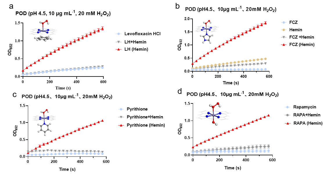
We systematically evaluated the POD activity of nanozymes formed by the conjugation of various antibiotics with hemin. The results demonstrated markedly enhanced POD activity across all tested nanozymes, validating the broad applicability and efficacy of this assembly strategy for multiple antibiotic classes.

Fig.2 POD-like activity of  Levofloxacin HCl(LH) (Hemin) (a)、Fluconazole (FCZ) (Hemin) (b)、pyrithione (Hemin) (c)、Rapamycin(RAPA) (Hemin) (d)
Fig.1 POD-like activity of Levofloxacin HCl(LH) (Hemin) (a)、Fluconazole (FCZ) (Hemin) (b)、pyrithione (Hemin) (c)、Rapamycin(RAPA) (Hemin) (d)

In Vitro Antimicrobial Activity

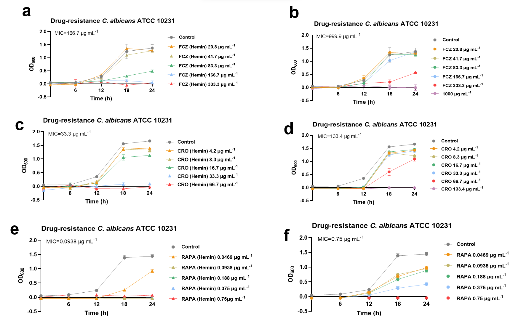
Antibacterial efficacy of the synthesized nanozymes was evaluated to determine whether the assembly strategy enhances bactericidal activity and broad-spectrum applicability. All antibiotic–hemin co-assemblies exhibited significantly stronger antimicrobial effects compared to their individual components.

Fig.3 Anti-C. albicans activity of FCZ (Hemin)、CRO(Hemin)、RAPA (Hemin)
Fig.2 Anti-C. albicans activity of FCZ (Hemin)、CRO(Hemin)、RAPA (Hemin)

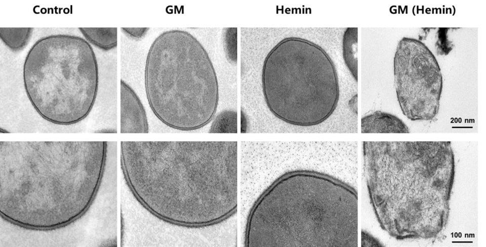
The synergistic effect of ferrous protoporphyrin in promoting the anti-MRSA activity of gentamicin (GM)

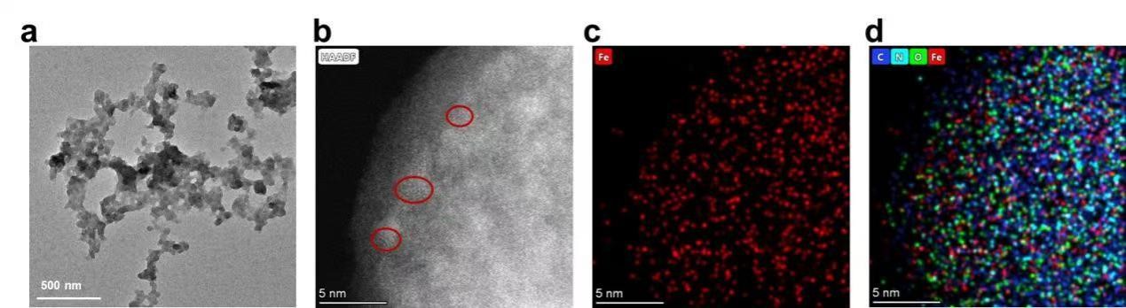
TEM Characterization of GM (Hemin)

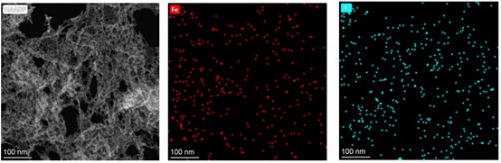
TEM revealed a cross-linked spherical nanostructure for the GM (Hemin) (Fig. 8a). High-angle annular dark-field imaging (Fig. 8b) showed uniform iron distribution at 5 nm resolution, while energy-dispersive X-ray spectroscopy(EDS) (Fig.8c,d) confirmed homogeneous distribution of Fe, C, N, and O elements throughout the assembly, verifying successful incorporation of hemin-derived iron.

Fig.7 TEM images (a), High-Angle Annular Dark-Field image (b), and EDS (c: Iron; d: Carbon, Nitrogen, Oxygen, Iron) of GM(Hemin)
Fig.3 TEM images (a), High-Angle Annular Dark-Field image (b), and EDS (c: Iron; d: Carbon, Nitrogen, Oxygen, Iron) of GM(Hemin)

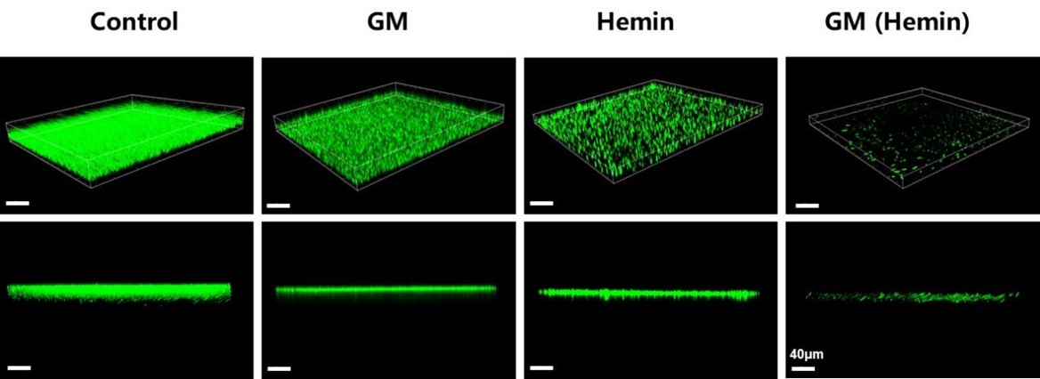
CLSM for Biofilm Eradication Assessment

Three-dimensional confocal laser scanning microscopy revealed significant reduction in bacterial density and biofilm thickness following gentamicin nanozyme treatment, providing direct visual evidence of biofilm disruption capability consistent with quantitative plate counting results.

Fig.9 The scavenging effect of GM (Hemin) on Methicillin-Resistant Staphylococcus aureus (MRSA) biofilm was observed by laser confocal imaging.
Fig.4 The scavenging effect of GM (Hemin) on Methicillin-Resistant Staphylococcus aureus (MRSA) biofilm was observed by laser confocal imaging.

TEM Analysis of MRSA

TEM images of MRSA ultrastructure showed control cells maintaining regular, turgid morphology with intact cell walls and membranes. In contrast, gentamicin nanozyme-treated bacteria exhibited severe cellular shrinkage, membrane disruption, uneven cytoplasmic density, and evident intracellular content leakage.

Fig.10 Observation of MRSA treated with GM (Hemin) by transmission electron microscope.
Fig.5 Observation of MRSA treated with GM (Hemin) by transmission electron microscope.

The synergistic effect of ferrous protoporphyrin in promoting the antifungal activity of Fluconazole (FCZ)

SEM Characterization of FCZ(Hemin)

SEM characterization confirmed nanoscale dimensions of the co-assembled fluconazole nanozyme, with EDS mapping showing homogeneous distribution of characteristic Fe and F elements, providing direct evidence of successful hemin-fluconazole co-assembly.

Fig.14 SEM image and element energy spectrum analysis (Fe and F) of FCZ (Hemin)
Fig.6 SEM image and element energy spectrum analysis (Fe and F) of FCZ (Hemin)

SEM Analysis of C. albicans

SEM observations revealed that the control C. albicans cells exhibited a plump and intact morphology. In contrast, the cells treated with the fluconazole nanozyme displayed a collapsed and shrunken surface with evident pores.

Fig.16 SEM images of C. albicans treated with FCZ (Hemin).
Fig.7 SEM images of C. albicans treated with FCZ (Hemin).