Antibiotic nanozyme synthesis
Background
Antibiotics contain diverse functional groups—such as imidazole, amino, hydroxyl, phenyl, and pyridine moieties—that can effectively displace chloride ions in hemin and serve as axial ligands to the Fe–N4 planar center, forming a pentacoordinated structure. These units can further self-assemble into nanoaggregates through various non-covalent interactions, including hydrogen bonding, salt bridges, π–π stacking, and cation-π interactions, ultimately leading to the co-assembly of antibiotic-integrated nanozymes.
Material and Methods
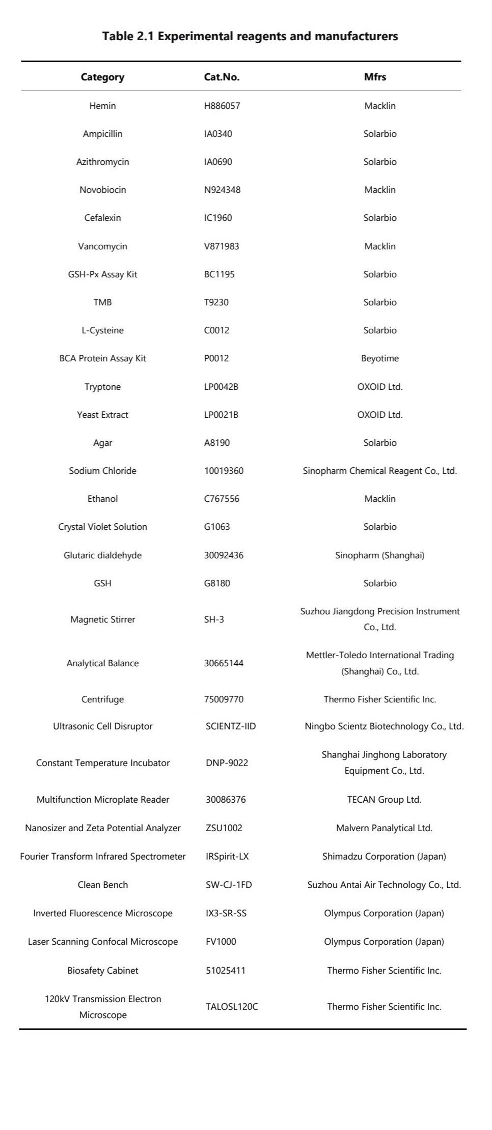
Materials:Hemin,Cefalexin(CFX),Gentamicin(GM),Rapamycin(RAPA), Ceftriaxone Sodium(CRO),Fluconazole(FCZ),sodium hydroxide (NaOH),dimethyl sulfoxide (DMSO)
Procedure
Hemin and gentamicin(or or alternatively, CFX,GM,RAPA,CRO,FCZ,etc.) were dissolved in a specified molar ratio under continuous stirring. The resulting mixture was then subjected to a drying process to obtain the final antibiotic nanozyme product.
Characterisation
Scanning Electron Microscopy (SEM) Characterization of Antibiotic Nanozyme
The antibiotic nanozyme was prepared at an appropriate concentration, dispersed by sonication at 100 W for 10 min, and then drop-cast onto a smooth silicon wafer attached to a sample stub. After drying in an oven, the sample was sputter-coated with a gold layer for 30 s using an ion sputterer before surface morphology characterization by scanning electron microscopy (SEM).
Transmission Electron Microscopy (TEM) Characterization of Antibiotic Nanozyme
A 10 μL aliquot of the uniformly dispersed antibiotic nanozyme aqueous solution was carefully pipetted onto a copper grid support film. The sample was allowed to settle at room temperature for 20 minutes, after which excess liquid was gently removed from the edge of the support film using absorbent filter paper. The grid was subsequently air-dried at room temperature and examined under a 120 kV transmission electron microscope for morphological observation. High-resolution transmission electron microscopy (HRTEM) was employed for dark-field imaging, and energy-dispersive X-ray spectroscopy (EDS) was utilized for elemental mapping analysis.
Peroxidase-like Activity Assay
Peroxidase-like activity of antibiotic nanozymes (including Gentamicin-,Rapamycin-, Ceftriaxone- and Fluconazole-based nanozymes) was assessed using 3,3′,5,5′-tetramethylbenzidine (TMB) as the substrate. The detailed procedure was as follows:
i. A 20 mg/mL TMB stock solution was prepared in dimethyl sulfoxide (DMSO) and stored protected from light.
ii. A 0.2 M acetate buffer (pH 4.5) was prepared.
iii. Antibiotic nanozyme solutions were prepared at 100 μg/mL in ultrapure water, followed by sonication at 100 W for 10 min. Control solutions of hemin chloride and the corresponding antibiotics at the same concentration were also prepared.
iv. The reaction was carried out in a 96-well plate with a total volume of 100 μL per well, containing 10 μL of gentamicin nanozyme or control component, 1 μL of TMB solution, 0.2 μL of 10 M H₂O₂, and 88.8 μL of 0.2 M acetate buffer (pH 4.5).
v. The absorbance at 652 nm was measured every 30 s for 10 min using a multifunctional microplate reader to monitor peroxidase-like activity.
Determination of Antimicrobial Activity of Antibiotic Nanozyme in vitro
The antifungal activity
i. C. albicans stored at -80°C was thawed, inoculated into 10 mL SDB liquid medium and incubated overnight in an air incubator at 37 °C with oscillations. The following day, the bacterial suspension was transferred to fresh medium and cultured for an additional 6-7 h at 37°C to activate the fungus,ensuring it reached the logarithmic growth phase.
ii. the initial inoculum concentration of the C. albicans suspension was approximately 10⁵-10⁶ CFU mL⁻¹, and the medium volume was 10 mL. To each well, 100 µL of C. albicans suspension and 100 µL of FCZ (Hemin) solution (or RAPA (Hemin), CRO (Hemin), or other components) at different concentrations were added to 800 µL of SDB medium and mixed thoroughly. As controls, the corresponding resistant antibiotics (FCZ, ITZ, or 5-FC) were added in the same volume (100 µL) along with 100 µL of bacterial solution and 800 µL of SDB medium as the negative control. The blank control consisted of 100 µL of bacterial solution and 900 µL of SDB medium. All groups were cultured at 37°C with shaking in an incubator for 24 h. After incubation, 200 µL of the suspension from each group was transferred to a microplate, and the absorbance was measured at 600 nm using a microplate reader.
The antibacterial activity
The antibacterial activity of GM (Hemin) against MRSA (ATCC43300) was determined using the same experimental method as for C. albicans.
i. The MRSA strains, stored at -80°C, were thawed and inoculated into 5 mL of LB liquid medium, followed by incubation with shaking at 37°C overnight. The next day, the bacterial solution was transferred to fresh LB medium at a 1:100 dilution and incubated at 37°C for approximately 2 h to activate the bacteria and allow them to enter the logarithmic growth phase, with a bacterial concentration of approximately 8×10⁸ CFU mL-1.
ii. 100 µL of MRSA suspension and 100 µL of GM (Hemin) solution (or other components) were added to 800 µL of LB liquid medium. The bacterial cultures were then incubated at 37°C for 24 h with continuous shaking. The absorbance at 600 nm of each group was measured using a multifunctional microplate reader to assess the antibacterial effect.
TEM for MRSA
i. An overnight culture of MRSA was subcultured and activated for 4 hours until it reached the mid-logarithmic phase (approximately 1 × 10⁹ CFU/mL).
ii. One-milliliter aliquots of the bacterial suspension were centrifuged in separate tubes, and the supernatant was discarded. The bacterial pellets were resuspended in 1 mg/mL solutions of either the gentamicin nanozyme or its individual components, followed by incubation at 37°C for 12 hours.
iii. After incubation, the bacteria were washed twice with phosphate-buffered saline (PBS). The pellets were then fixed with 1 mL of 2.5% glutaraldehyde and stored at 4°C for a minimum of 2 hours.
iv. The fixed bacterial samples were centrifuged and washed three times with ddH₂O. Subsequently, they were subjected to sequential dehydration in a graded ethanol series (50%, 70%, 85%, 95%, and 100%). After critical point drying, the specimens were sputter-coated and imaged using a scanning electron microscope to examine the surface morphology.
v. For TEM observation, the fixed samples were embedded in resin and ultrathin-sectioned. The sections were then placed on copper grids and observed under a transmission electron microscope to assess the internal structure and morphology.
SEM for C. albicans
The bacterial suspension cultured overnight was transferred and activated for 6h. A 1 mL aliquot of the fungal suspension was taken from each centrifuge tube, centrifuged, and the supernatant was discarded. The drug-resistant working suspension was then treated with FCZ (Hemin) (at a final concentration of 1 mg mL-1), while the drug-resistant bacteria treated with FCZ and Hemin were used as the control group. The samples were fixed with 4% paraformaldehyde, followed by scanning electron microscopy (SEM) imaging.
Investigation of Antimicrobial Mechanisms
Confocal Laser Scanning Microscopy (CLSM) for Biofilm Eradication Assessment
i. Overnight bacterial culture was subcultured and activated for 4 h to approximately 1×10⁹ CFU/mL for subsequent use.
ii. A 24-well plate was prepared with cell climbing slides, and 1 mL of bacterial suspension was added to each well. The plate was incubated at 37°C for 24 h to allow biofilm formation.
The medium was carefully aspirated, and 1 mL of 100 μg/mL gentamicin nanozyme or control solutions was gently added along the well wall. The plate was incubated at 37°C for 3 h.
iv. After treatment, the liquid was removed, and biofilms were stained with 0.5 μM SYTO 9 solution at 37°C in the dark for 30 min. The slides were then washed with PBS and carefully retrieved.
v. Each slide was mounted inverted on a clean glass slide with 5 μL of anti-fade mounting medium and imaged using confocal laser scanning microscopy.